Interactions between sensory and emotions
Beautiful art, plus- Transformation of sensory cues into negative #emotion. Xiong, Zhang, Tao & colleagues https://t.co/8obcJrrmmZ pic.twitter.com/zEgCePPaac
— Neuron (@NeuroCellPress) September 12, 2018
Mouse medial preoptic area (mPOA) glutamatergic neurons encode extremely negative valence, promote anxiety-like behaviors, and inhibit parental behavior; mPOA GABAergic neurons have the opposite effectshttps://t.co/O6o3vlfIY2
— Nature Neuroscience (@NatureNeuro) February 2, 2021
Emotions evoked by environmental cues are essential for animal survival and life quality. However, how different sensory stimuli evoke different emotional reactions? For example, when we hear a loud noise, we may feel uncomfortable and avoid it. In other words, the loud noise induces an emotional effect -- aversion and a motional effect -- avoidance. But how and where this transformation from sensory cues to emotions and motional behaviors happens?
Among those emotions, anxiety, a representation of negative emotion, helps us keep vigilance. However, overly displayed anxiety may impair our life quality as well. Due to the increasing pressures of life and work in modern society, anxiety disorders are becoming more prevalent and represent a significant societal challenge. A thorough understanding of the neural circuits governing this emotional state is necessary to improve existing or develop new treatment strategies for anxiety disorders. Although anxiety is known to be controlled by distributed brain networks, key components for its initiation, maintenance, and coordination with behavioral state remain poorly understood.
In this project, we aim to decipher the mystery about interactions between sensory and emotion, especially for aversion and anxiety.
Techniques
Research highlights
1. A Non-canonical Central Auditory Pathway Contributes to Fear Conditioning
Firstly, starting from auditory sensory processing pathway, we have discovered a previously unrecognized central auditory pathway, which is involved in processing aversive acoustic signals and contributes to auditory fear conditioning. It opens a window to reconsider the overall strategy for auditory information processing in the brain.
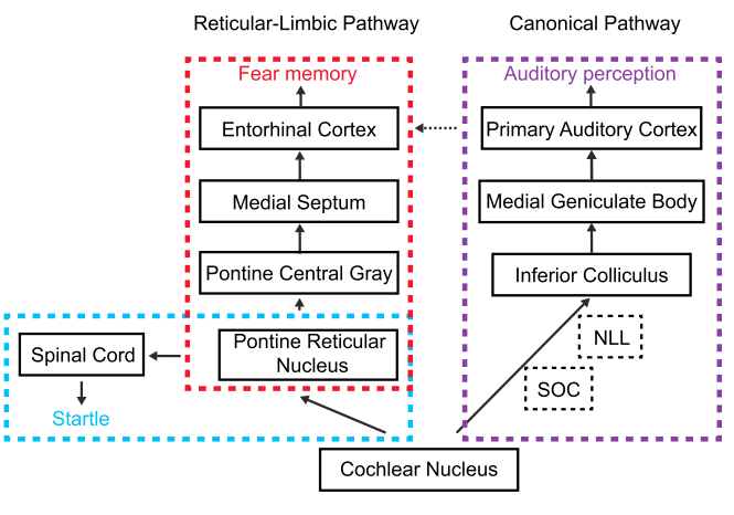
Reticular-Limbic and Canonical Lemniscal Pathways
In awake mice, we found that auditory responses in entorhinal cortex (EC) cannot be explained by a previously proposed relay from auditory cortex (AC) based on response properties. By combining anatomical tracing and optogenetic/pharmacological manipulations, we discovered that EC received auditory input primarily from the medial septum (MS), rather than AC. A previously uncharacterized auditory pathway was then revealed: it branched from the cochlear nucleus (CN), and via caudal pontine reticular nucleus (PRN), pontine central gray (PCG), and MS, reached EC. Neurons along this non-canonical auditory pathway responded selectively to high-intensity broadband noise, but not pure tones. Disruption of the pathway resulted in an impairment of specifically noise-cued fear conditioning. This reticular-limbic pathway may thus function in processing aversive acoustic signals.
2. A Septal-Habenular Pathway for Transforming Sensory Cues into Aversive Emotion
Next, we asked how the sensory cues transformed into both emotion and behavior reactions. We have revealed a circuit for processing innately aversive sensory signals, with glutamatergic projections from a nucleus in basal forebrain -- MS to two distinct downstream: lateral habenula (LHb) in thalamus and pre-optic area (POA) in hyperthalamus to generate the emotional and a motional effect, respectively. This role of MS in mediating aversion is previously unrecognized.
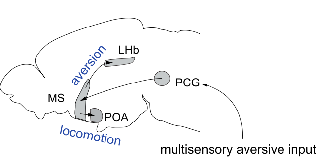
A bottom-up multisensory neural pathway from PCG to MS and then to LHb (which mediates aversion) and POA (which mediates enhancement of locomotion).
we found that MS mediates aversion induced by both auditory and somatosensory stimuli. Ablation of glutamatergic or GABAergic MS neurons results in impaired or strengthened aversion, respectively. Optogenetic activation of the two cell types results in place avoidance and preference, respectively. Cell-type-specific screening reveals that glutamatergic MS projections to the LHb are responsible for the induction of aversion, which can be antagonized by GABAergic MS projections to LHb. Additionally, the sensory-induced place avoidance is facilitated by enhanced locomotion mediated by glutamatergic MS projections to the POA. Thus, MS can transmit innately aversive signals via a bottom-up multimodal sensory pathway and produce concurrent emotional and motional effects, allowing animals to efficiently avoid unfavorable environments.
3. A Key Brain Area for coordinating anxiety and parental behavior
Furthermore, we moved forward to the interactions between emotion and social behavior, and focused on anxiety emotion. We found that a area in hyperthalamus -- medial preoptic area (mPOA) -- plays a key role in mediating stress-induced anxiety and parental behavior. The aglutamatergic neurons in mPOA encode extremely negative valence, promote anxiety-like behaviors, and inhibit parental behavior; mPOA GABAergic neurons have the opposite effects.
Glutamatergic neurons in mPOA encode an extremely negative feeling that the mice prefer to stay in the water/heat/electric shock side than receiving the optogenetic stimulation.
The anxiogenic stressors, such as forced swimming, a heat plate and electric shocks, elicit acute and prolonged responses in glutamatergic neurons of the mouse mPOA. These neurons encode extremely negative valence and mediate the induction and expression of anxiety-like behaviors. Conversely, mPOA GABA-containing neurons encode positive valence and produce anxiolytic effects. Such opposing roles are mediated by competing local interactions and long-range projections of neurons to the periaqueductal gray (PAG). The two neuronal populations antagonistically regulate anxiety-like and parental behaviors: anxiety is reduced, while parenting is enhanced and vice versa. Thus, by evaluating negative and positive valences through distinct but interacting circuits, the mPOA coordinates emotional state and social behavior.
Related Publications